Vendor-agnostic multiplex image analysis right here in your web browser. World-class segmentation and registration. Neuroscience toolkit. For multi-channel Immunofluorescence, spatial proteomic, transcriptomics and more.
Sign Up

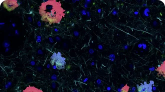
World-class cell segmentation.

Also for cells with complex morphologies such as neurons and glia. Supports CD45, NeuN, Iba1, GFAP, DAPI and many more.

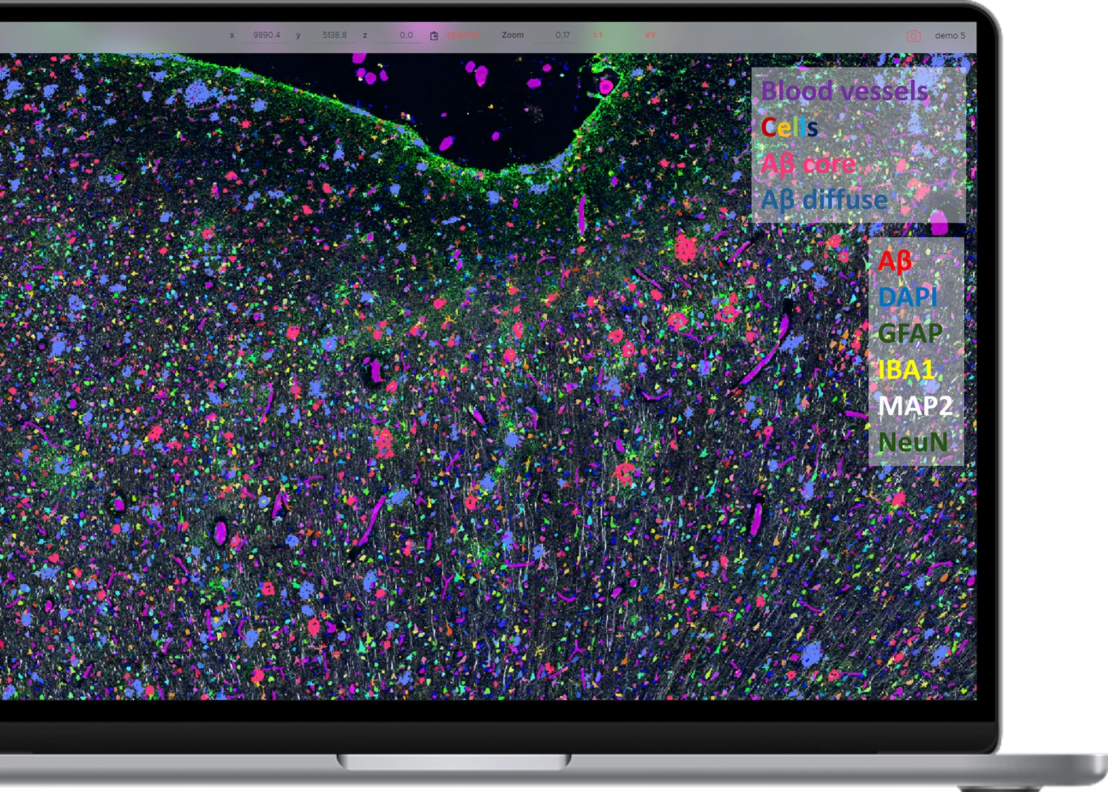
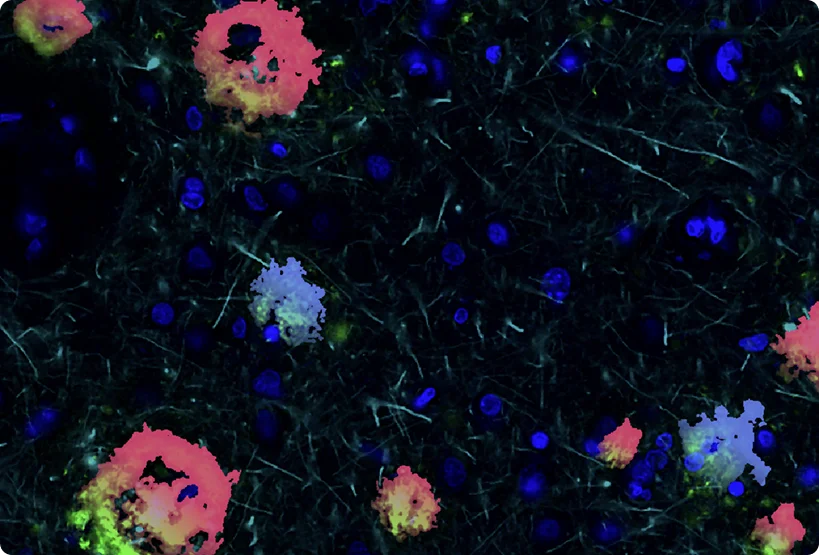
Neuropathology analysis.

Classify pathological protein aggregates of Amyloid β, pTau, α-Synuclein, TDP43 and more.
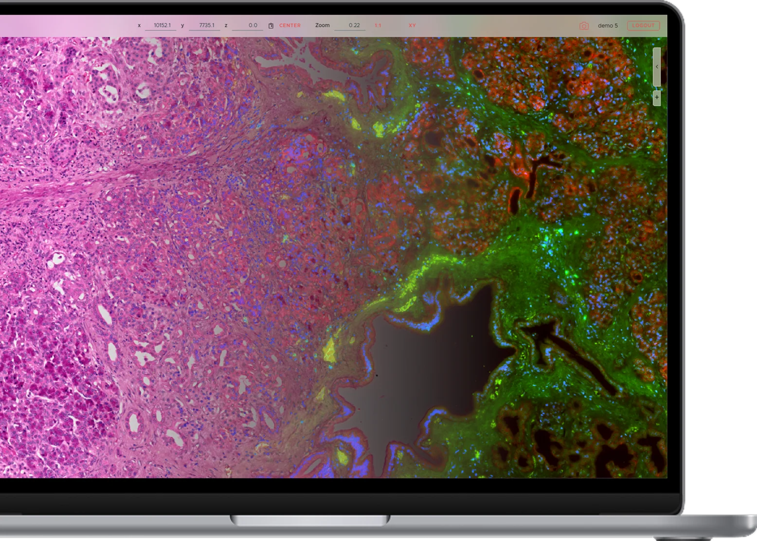
Segment H&E images.

Leverage well-established IHC protocols, multiplexed.

Elastic registration.

Correct complex deformations accross consecutive staining cycles, protocols or physical sections.
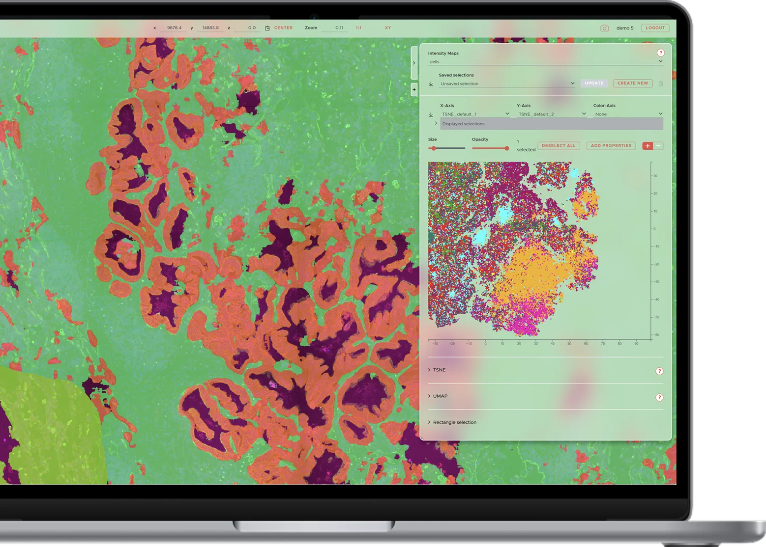
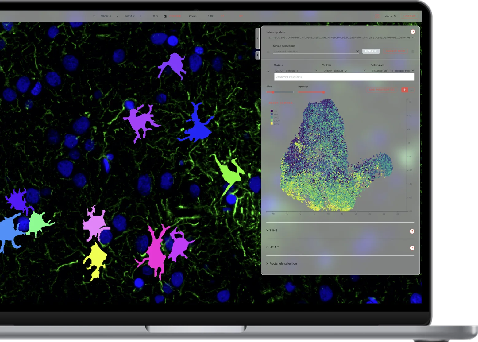
Detailed segmentation from fluorescent markers such as NeuN, IBA1, GFAP, CD45 and more enables precise marker mapping and morphometric quantification.

The elastic alignment workflow allows you to integrate different staining protocols (e.g., spatial proteomics and spatial transcriptomics) on the same section or accross consecutive physical sections.

Leverage a flexible combination of manual gating, automated cell clustering, and marker-independent tissue segmentation to group cells into biologically meaningful phenotypes. Analyze and compare these clusters across conditions, regions, or tissue types to uncover spatial patterns and functional insights.

Go beyond traditional single-cell analysis by identifying spatial biomarkers that capture the architecture and context of tissue. SPATIAL quantifies features such as cell shape, location, size and branching patterns — unlocking new classes of digital biomarkers that reflect molecular, morphological and spatial complexity.

Workflow

SPATIALTM provides access to an end-to-end image analysis workflow — from data upload to spatial biomarker discovery.

Start Now

applications

Comprehensive in silico tissue characterization in Alzheimer's and Parkinson's disease by spatial proteomics
Learn more
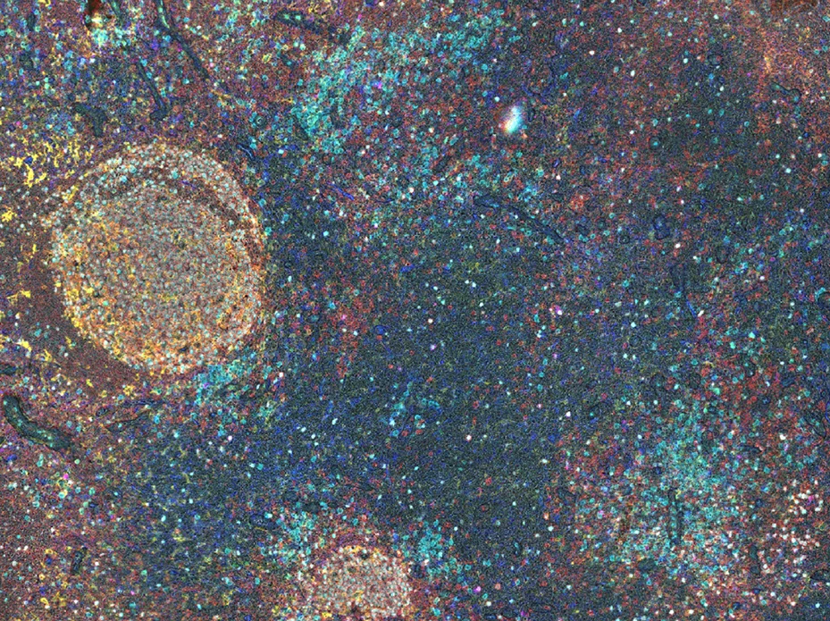
Webinar: High-plex immunohistochemistry - Spatial biology links patient survival with lymph node B cell responses in head & neck cancer
Learn more
Start your free trial

Start analyzing data today

Sign Up

Frequently asked questions

Everything you need to know about the product and billing.

Most common file types are supported, and we keep adding more. Currently supported file types include OME-TIFF, raw TIFF series, Zeiss CZI, Leica LIF and also include the datasets generated by Akoya Phenocycler, Bruker Spatial Biology CosMx and CellScape, Lunaphore COMET, 10xGenomics Visium, VisiumHD, Xenium and Vizgen MERSCOPE.

SPATIALTM can integrate stacks of dense images (e.g., IHC or IF image series) and sparse images (e.g., lists of coordinates generated by mISH). This makes it broadly compatible with analysis workflows in spatial proteomics, transcriptomics and metabolomics. It's confirmed to work well with Akoya Phenocycler, Bruker Spatial Biology CosMx and CellScape, Lunaphore COMET, 10xGenomics Visium, VisiumHD, Xenium and Vizgen MERSCOPE and more.

We provide comprehensive API access, allowing you to develop your own scripts to interface with SPATIALTM.

SPATIALTM is available through an affordable, simple and transparent pay-as-you-go model. We are happy to provide a custom quote for bulk orders or if you'd like to outsource the end-to-end analysis workflow to us.

SPATIAL works on any modern browser, leveraging cloud computing for processing. This means you can use our platform even on low-powered machines.

It's fully elastic, and scalable to arbitrary size. Some data can't be analyzed without this, but we've found that even nominally well-aligned data can profit from running our registration pipeline. Pixel-perfect precision is essential to get minimal cross-talk!

We segment cells using image channels that outline cellular morphology, optionally combined with a nucleus channel. The method works on any image channel on which cell outlines can be made out. For example, antibody-derived stains of CD45, NeuN, Iba1, GFAP, DAPI and many more can be used, but H&E or Hematoxylin staining works as well. Multiple markers can be combined for segmentation.

There is no set limit on dataset size. 2-dimensional datasets are billed per dataset; 3-dimensional datasets are billed by amount of data.

The fees are invoiced to institutions, payable via bank transfer.

Results are accessible through the SPATIAL browser interface and downloadable in standard formats through the user interface, or through an API for advanced users.

We take data security very seriously. All data provided by our customers is kept strictly confidential and maintained on secure, EU-based and ISO 27001-compliant infrastructure.

For more information, schedule a personalized remote demo right here on this website! We're also happy to arrange an on-site visit if we're in your area.

Latest News